|
ULTRASOUND OF
PARVOVIRUS B19 INFECTION |
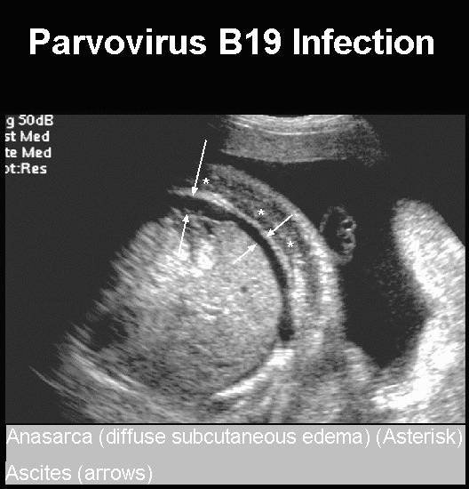
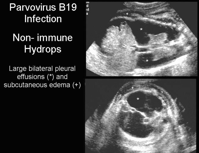
- Nonimmune fetal hydrops.
- Subcutaneous edema.
- Ascites.
- Pleural and pericardial effusions.
- May resolve spontaneously with good outcome (1).
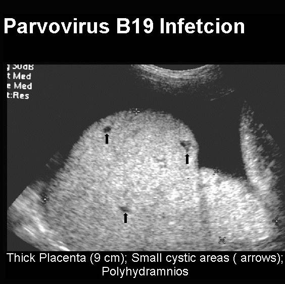
- Polyhydramnios (2).
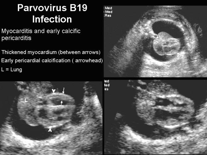
- Myocarditis / Cardiomyopathy / Pericarditis.
- Thick placenta (due to the hydrops).
- No structural fetal anomalies. It appears that the frequency of congenital malformation after infection is not increased as compared with the general population (1).
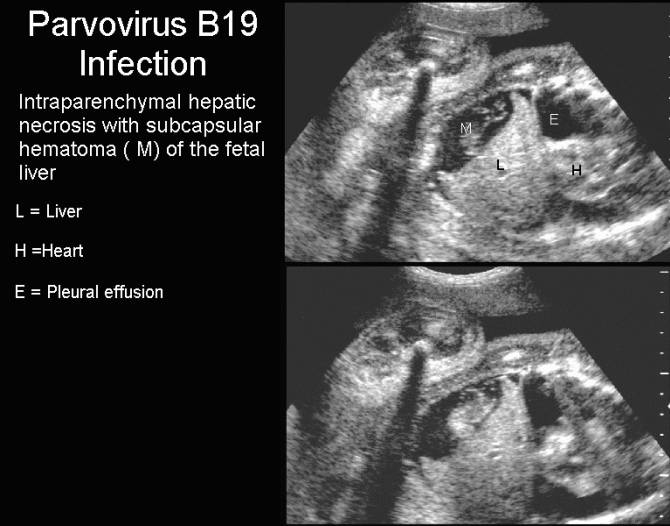
- Hepatic fibrosis and necrosis (either due to direct effect on hepatocytes or indirect damage from hemosiderin deposition in hepatocytes (3).
- Symmetric IUGR (2). The fetal growth appears to catch up once the hydrops resolves (4).
- Assessment of the degree of fetal anemia –
MC- PSV.
|
|
|
|
|
|
|
|
|
|
|
|
Parvovirus – Pleural
effusions and anasarca Parvovirus – Reverse
diastolic flow in the umbilical artery
|
|
|
|
|
|

REFERENCES |
- Torok TJ, Wang Q-Y, Gary GW Jr et.al. Prenatal diagnosis of intrauterine infection with parvovirus B19 by the polymerase chain reaction technique. Clin Infect Dis 1992;14:149-155.
- Bhal PS,
- Kilham L, Margolis G. Problems of human concern arising from animal models of intrauterine and neonatal infections due to viruses. Prog Med Virol 1975;20:113.
- Pryde PG, Nugent CE, Pridjian G et.al. Spontaneous resolution of non-immune hydrops secondary to parvovirus B19 infection. Obstet Gynecol 1992;79:859-861.